
Models Forced Expiratory Volume procedure using the Maxwell one chamber viscoelastic compartment model taken from Bates (ch. 7)
Description
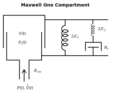
	This model adapts the Maxwell single compartment viscoelastic lung as
developed by Bates to the forced expiratory volume (FEV) procedure. The model
includes a resistance term (represented by airway resistance) and a spring recoil
term (spring resistance represented by 1 / lung compliance, 1/C) for the lung and
lung wall. The combination of spring and dashpot in series, a Maxwell body, accounts
for the viscoelastic behavior of this model. Several commonly used lung volumes have
been calculated and reported. This model can be modified to add to a larger model or
can be used alone as a simple model. Several data sets have been added and the model
has been fit to each.
	This model has several problems. First, the driving pressure of the system is
external, not from the chest wall, and therefore acts like a respirator pumping up
the lung with positive pressure and sucking the air out with negative pressure, the
reverse of normal ventilation. This is the opposite of the "forced expiration"
compressing the chest and raising intrapleural pressure. However the mechanics of
the lung are reasonably modeled, giving a 2-component outflow that can be fitted to
actual data.
	The second problem is that the compliances, C1 and C2, are constant instead
of being functions of the volume. In reality the FEV ends, at Vlung = Functional
Reserve Capacity - Expiratory Reserve Volume = Residual Volume, when the force of
chest muscle contraction cannot make the chest smaller (equivalent to compressing the
spring further). The passive pressure-volumes curves should be concave upward for
both chest and lungs at volumes above FRC, At FRC the chest recoil is toward
enlargement and the lung recoil is toward shrinkage, so the combination produces a
negative intrapleural pressure at rest.
	Thirdly, air is assumed to be incompressible. In reality air flow inward
through the mouth is less than flow into the lungs because of the additional volume
contributed by water vapor pressure rising to 47 mmHg. With an FEV the volume is
compressed, often by 1/8th, i.e. 100 mmHg exhalation pressure added to 760 mmHg.
	This model differs from the FEV_simple model (insert model #) in its addition
of viscoelastic behavior created by the Maxwell body representing chest wall
mechanics. By adding a second spring and resistor set, this model is capable of
fitting well to FEV data curves.
Figure
Equations
The equations for this model may be viewed by running the JSim model applet and clicking on the Source tab at the bottom left of JSim's Run Time graphical user interface. The equations are written in JSim's Mathematical Modeling Language (MML). See the Introduction to MML and the MML Reference Manual. Additional documentation for MML can be found by using the search option at the Physiome home page.
- Download JSim model MML code (text):
- Download translated SBML version of model (if available):
- No SBML translation currently available.
- Information on SBML conversion in JSim
We welcome comments and feedback for this model. Please use the button below to send comments:
J.H.T. Bates. Lung Mechanics: An Inverse Modeling Approach. Cambridge University
Press, (2009)  Chapter 3: 37-44, Chapter 7: 117-118.
Please cite https://www.imagwiki.nibib.nih.gov/physiome in any publication for which this software is used and send one reprint to the address given below:
The National Simulation Resource, Director J. B. Bassingthwaighte, Department of Bioengineering, University of Washington, Seattle WA 98195-5061.
Model development and archiving support at https://www.imagwiki.nibib.nih.gov/physiome provided by the following grants: NIH U01HL122199 Analyzing the Cardiac Power Grid, 09/15/2015 - 05/31/2020, NIH/NIBIB BE08407 Software Integration, JSim and SBW 6/1/09-5/31/13; NIH/NHLBI T15 HL88516-01 Modeling for Heart, Lung and Blood: From Cell to Organ, 4/1/07-3/31/11; NSF BES-0506477 Adaptive Multi-Scale Model Simulation, 8/15/05-7/31/08; NIH/NHLBI R01 HL073598 Core 3: 3D Imaging and Computer Modeling of the Respiratory Tract, 9/1/04-8/31/09; as well as prior support from NIH/NCRR P41 RR01243 Simulation Resource in Circulatory Mass Transport and Exchange, 12/1/1980-11/30/01 and NIH/NIBIB R01 EB001973 JSim: A Simulation Analysis Platform, 3/1/02-2/28/07.