
The autoregulatory device (ARD) developed by Gao et al. is reproduced here to simulate the autoregulation of cerebral blood flow in the 300 to 50 micron diameter vessels.
Description
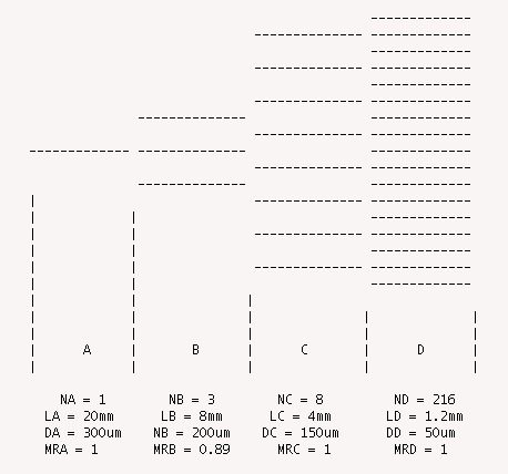
  This model represents the autoregulation of blood flow through a portion
  of the cerebral microvasculature as developed by Gao et al. (Am J Physiol
  274:H1023-H1031, 1998).  The microvasculature is described by a
  representative segment model where each of the four vascular regions are
  described by a specified number of identical vessel segments.  In the first
  region with a control diameter of 300 microns there is a single segment.
  In the second region with a control diameter of 200 microns there are 3
  segments.  The third region with a control diameter of 150 microns has 8
  segments while the final region with a control diameter of 50 microns has
  216 segments.

  The number of segments in each region is determined in such a manner that
  Ni*Di^3 is relatively constant throughout the network.  This is based
  on Murray's cost analysis of network branching morphology (PNAS 12:207-214,
  1926). The vessel segments in each region are then allowed to vary as a
  function of the  systemic mean arterial pressure (MAP) according to
  empirical fits to the in vivo data of Kontos et al. (Am J Physiol 234:
  H371-H383, 1978).

  It should be noted that the empirical expression used to fit the Kontos et
  al. data, Equation 3, is incorrect in the published paper. See Equations
  section below.

  The parameters from Table 3 in the paper reproduce the diameter versus
  pressure curves shown in Figure 5 with the equation quoted below.
where MR is the Murray's law ratio: Nx*Rx^3 / NA*RA^3.
Equations
The autoregulatory device developed by Gao uses an empirical fit to data from Kontos et al. where the equation for the variation in diameter di as a function of the systemic
where each compartment, i, of the autoregulatory device (i = A through D) has a different regulatory response based on the set of fit parameters, a0i to a7i . The resistances in each compartment can then be calculated from the diameter of the segments using the following expression:
where Ri is the resistance in compartment i,  is the viscosity of blood, Li is the segment length in compartment i, Ni is the number of segments in compartment i and di is the segment diameter determined from the empirical fit above.
The resistances in each compartment are then added to get the total resistance of the ARD and then the flowrate, Q, and effective diameter, deff, can then be calculated from the following expressions:
The equations for this model may be viewed by running the JSim model applet and clicking on the Source tab at the bottom left of JSim's Run Time graphical user interface. The equations are written in JSim's Mathematical Modeling Language (MML). See the Introduction to MML and the MML Reference Manual. Additional documentation for MML can be found by using the search option at the Physiome home page.
- Download JSim model MML code (text):
- Download translated SBML version of model (if available):
We welcome comments and feedback for this model. Please use the button below to send comments:
 Gao E, Young WL, Pile-Spellman J, Ornstein E and Ma Q.
 Mathematical considerations for modeling cerebral blood flow autoregulation to systemic arterial pressure.
 American Journal of Physiology, Heart and Circulatory Physiology 274:H1023-H1031, 1998.

 Kontos HA, Wei EP, Navari RM, Levasseur JE, Rosenblum WI and Patterson JL.
 Responses of cerebral arteries and arterioles to acute hypotension and hypertension.
 American Journal of Physiology, Heart and Circulatory Physiology 234:H371-H383, 1978.

 Murray CD; The physiological principle of minimum work. I. The vascular system and cost of blood volume.
 Proceedings of the National Academy of Sciences of the United States of America 12:207-214, 1926.
Please cite https://www.imagwiki.nibib.nih.gov/physiome in any publication for which this software is used and send one reprint to the address given below:
The National Simulation Resource, Director J. B. Bassingthwaighte, Department of Bioengineering, University of Washington, Seattle WA 98195-5061.
Model development and archiving support at https://www.imagwiki.nibib.nih.gov/physiome provided by the following grants: NIH U01HL122199 Analyzing the Cardiac Power Grid, 09/15/2015 - 05/31/2020, NIH/NIBIB BE08407 Software Integration, JSim and SBW 6/1/09-5/31/13; NIH/NHLBI T15 HL88516-01 Modeling for Heart, Lung and Blood: From Cell to Organ, 4/1/07-3/31/11; NSF BES-0506477 Adaptive Multi-Scale Model Simulation, 8/15/05-7/31/08; NIH/NHLBI R01 HL073598 Core 3: 3D Imaging and Computer Modeling of the Respiratory Tract, 9/1/04-8/31/09; as well as prior support from NIH/NCRR P41 RR01243 Simulation Resource in Circulatory Mass Transport and Exchange, 12/1/1980-11/30/01 and NIH/NIBIB R01 EB001973 JSim: A Simulation Analysis Platform, 3/1/02-2/28/07.