
Simple compartmental model including soluble gas transport in the alveoli, transfer between the bronchial circulation and the conducting airways, and metabolism.
Description
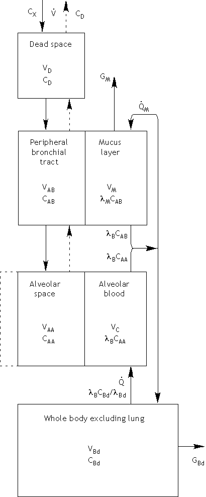
  This is a simple model of the pulmonary system where the lung is subdivided into
  compartments representing air spaces: dead space, peripheral bronchial tract, and
  the alveolar space; blood volumes: upper airway mucus, pulmonary capillary, systemic
  blood flow; and tissue space: whole body excluding the lungs. The upper airway mucus
  layer resides along the peripheral bronchial tract and has a continuous blood flow
  QM which effectively communicates with the gas in the bronchial tract.
Equations
-
Ventilation
The alveolar compartment changes volume with ventilation. Prior to steady state, the lung undergoes tidal breathing, represented by a sinusoid
After a pre-determined number of breaths, a secondary maneuver is performed: prolonged exhale or rebreathing. For a prolonged exhalation maneuver, the lungs are brought quickly to total lung capacity (TLC), then drained at a constant rate to the residual volume (RV).

In a rebreathing maneuver, an external bag is attached and the subjects breaths a number of breaths, generally 8, directly into and out of a bag.

Compartmental Definitions The following compartments use conservation of mass equations to follow the concentration of the soluble gas tracer.
-
Dead Space
-
Peripheral Bronchial Compartment
-
Alveolar Compartment

-
Body Compartment

-
Rebreathing Bag
- Download JSim model MML code (text):
- Download translated SBML version of model (if available):
We welcome comments and feedback for this model. Please use the button below to send comments:
  Kumagai S, and Matsunaga I. A lung model describing uptake of organic solvents and
  roles of mucosal blood flow and metabolism in the bronchioles. Inhal Toxicol 12: 491-510, 2000.
Please cite https://www.imagwiki.nibib.nih.gov/physiome in any publication for which this software is used and send one reprint to the address given below:
The National Simulation Resource, Director J. B. Bassingthwaighte, Department of Bioengineering, University of Washington, Seattle WA 98195-5061.
Model development and archiving support at https://www.imagwiki.nibib.nih.gov/physiome provided by the following grants: NIH U01HL122199 Analyzing the Cardiac Power Grid, 09/15/2015 - 05/31/2020, NIH/NIBIB BE08407 Software Integration, JSim and SBW 6/1/09-5/31/13; NIH/NHLBI T15 HL88516-01 Modeling for Heart, Lung and Blood: From Cell to Organ, 4/1/07-3/31/11; NSF BES-0506477 Adaptive Multi-Scale Model Simulation, 8/15/05-7/31/08; NIH/NHLBI R01 HL073598 Core 3: 3D Imaging and Computer Modeling of the Respiratory Tract, 9/1/04-8/31/09; as well as prior support from NIH/NCRR P41 RR01243 Simulation Resource in Circulatory Mass Transport and Exchange, 12/1/1980-11/30/01 and NIH/NIBIB R01 EB001973 JSim: A Simulation Analysis Platform, 3/1/02-2/28/07.