
A theoretical model dealing with endocytosis, exocytosis and caveolae invagination, describing plasmalemma homeostasis during cell growth and division, was proposed. It considers transmembrane pressure, membrane tension and mechanosensitivity of membrane processes.
Description
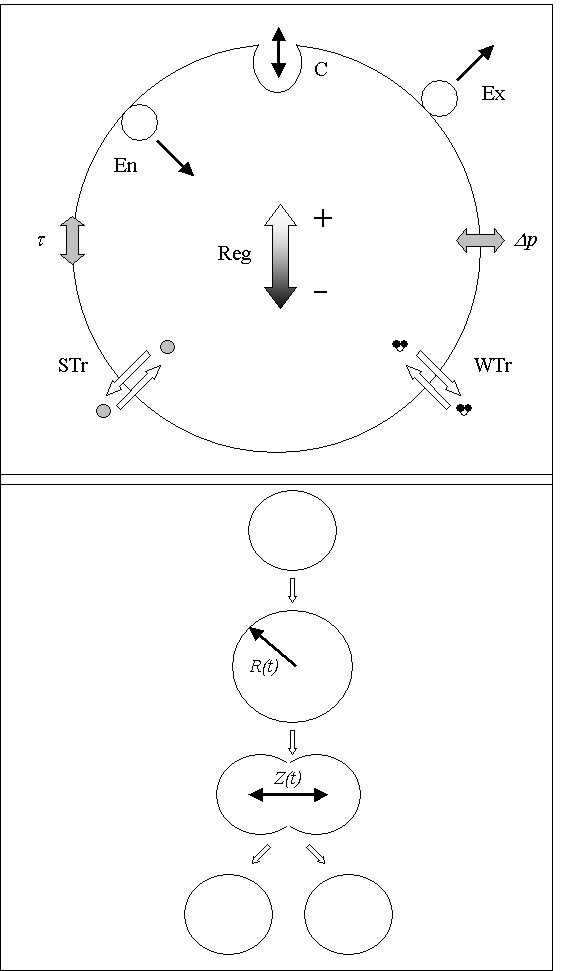
- A single cell was modeled as a mechanochemical set of plasmalemma, encapsulated cytoplasm and surrounding environment. It was assumed that membrane material can be delivered to the bilayer by exocytosis and unloaded by endocytosis. According to the model, locally flat membrane can invaginate forming bud-like caveolaes (C). Single cell volume can change due to the transport of water (WTr), small solute (STr), and endocytic (En) and exocytic (Ex) vesicles. The considered processes can be tension (t) sensitive. For simplicity the encapsulated volume as well as the excluded area of the membrane, occupied by membrane material of endocytic end exocytic vesicles, were not distinguished. Vesicle and the cell volume exceeding certain border limit were treated as incompressible. Additionally, the shape of the cell was assumed to be spherical, and characterized by time (t) dependent radius (R(t)).
- We assumed that a cell initiates mitotic separation when its volume reaches a certain minimum, and the membrane tension is not higher than a certain critical value. During mitosis the cell rearranges its geometry forming a two-kernel peanut shape, made of a symmetrical pair of cut and joined spheres, with the centers separated by a distance (Z(t)), shorter than the diameter of the sphere.
- The cell divides symmetrically when Z exceeds the distance 2R.

Figure: Dividing cell volume as a function of time.
Equations


where: t is time,
 is the hydraulic conductivity of the membrane,
 is excluded area of the cell membrane,
 is hydraulic pressure difference,
 is osmotic pressure difference,
 is the encapsulated volume of endocytic and exocytic vesicles,
 is the membrane excluded area of endocytic end exocytic vesicles,
 are the rates of endocytosis, exocytosis, budding and bud vanishing,
 is the number of buds (caveolaes).
- Download JSim model MML code (text):
- Download translated SBML version of model (if available):
We welcome comments and feedback for this model. Please use the button below to send comments:
Pawlowski PH, Mechanokinetic model of cell membrane: Theoretical analysis of plasmalemma homeostasis, growth and division. Journal of Theoretical Biology, Vol 249(1), 7 November 2007: 67-76.
Please cite https://www.imagwiki.nibib.nih.gov/physiome in any publication for which this software is used and send one reprint to the address given below:
The National Simulation Resource, Director J. B. Bassingthwaighte, Department of Bioengineering, University of Washington, Seattle WA 98195-5061.
Model development and archiving support at https://www.imagwiki.nibib.nih.gov/physiome provided by the following grants: NIH U01HL122199 Analyzing the Cardiac Power Grid, 09/15/2015 - 05/31/2020, NIH/NIBIB BE08407 Software Integration, JSim and SBW 6/1/09-5/31/13; NIH/NHLBI T15 HL88516-01 Modeling for Heart, Lung and Blood: From Cell to Organ, 4/1/07-3/31/11; NSF BES-0506477 Adaptive Multi-Scale Model Simulation, 8/15/05-7/31/08; NIH/NHLBI R01 HL073598 Core 3: 3D Imaging and Computer Modeling of the Respiratory Tract, 9/1/04-8/31/09; as well as prior support from NIH/NCRR P41 RR01243 Simulation Resource in Circulatory Mass Transport and Exchange, 12/1/1980-11/30/01 and NIH/NIBIB R01 EB001973 JSim: A Simulation Analysis Platform, 3/1/02-2/28/07.