
Description & purpose of resource
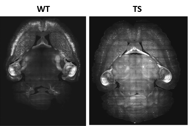
Whole brain images of a Down syndrome (DS) mouse model (Ts65Dn). The Thy1-eYFP-H was crossed with Ts65Dn mouse model for imaging excitatory neurons.The images of DS mice model (TS) and their wild type littermates (WT) were acquired with light sheet fluorescence microscopy (MuVi-SPIM, Luxendo), cleared with CLARITY. The whole brain dataset consist of 13 WT (8 males and 5 females) and 17 TS (11 males and 6 females). Magnification: 15x. Voxel resolution: 0.433um x 0.433um x2.00um.
Spatial scales
cellular
tissue
organ
This resource is currently
under early-stage development
DOI link to publication describing this resource
pending_to_be_publicly_available_at_BIL_repository
Link to resource
Keywords
whole brain image, Down syndrome mouse model, Ts65Dn, Thy1-eYFP-H
Table sorting checkbox
Off
Model type
topological
geometrical
Data type
expression
fluorescence
imaging