
(Matlab) Computational Model of Cellular Metabolic Dynamics in Skeletal Muscle Fibers during Moderate Intensity Exercise.
Description
(Abstract) Human skeletal muscles have different fiber types with distinct metabolic functions and physiological properties. The quantitative metabolic responses of muscle fibers to exercise provide essential information for understanding and modifying the regulatory mechanisms of skeletal muscle. Since in vivo data from skeletal muscle during exercise is limited, a computational, physiologically based model has been developed to quantify the dynamic metabolic responses of many key chemical species. This model distinguishes type I and II muscle fibers, which share the same blood supply. An underlying hypothesis is that the recruitment and metabolic activation of the two main types of muscle fibers differ depending on the pre-exercise state and exercise protocols. Here, activation measured by metabolic response (or enzymatic activation) in single fibers is considered linked but distinct from fiber recruitment characterized by the number (or mass) of each fiber type involved during a specific exercise. The model incorporates species transport processes between blood and muscle fibers and most of the important reactions/pathways in cytosol and mitochondria within each fiber type. Model simulations describe the dynamics of intracellular species concentrations and fluxes in muscle fibers during moderate intensity exercise according to various experimental protocols and conditions. This model is validated by comparing model simulations with experimental data in single muscle fibers and in whole muscle. Model simulations demonstrate that muscle-fiber recruitment and metabolic activation patterns in response to exercise produce significantly distinctive effects depending on the exercise conditions.
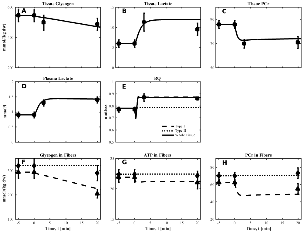
Figure: (from fig 3, Li et al. 2012) Simulated dynamics and experimental data for intracellular species concentrations in skeletal muscle in response to an increased work rate equivalent to cycle ergometer exercise at 50% VO2max for subjects corresponding to Experiment 1: (left to right) Glycogen, Lactate, Phosphocreatine (PCr) in tissue; Plasma Lactate, Respiratory quotient (RQ); Glycogen in fibers, and ATP in type I and II fibers; The lines represent model simulations of whole muscle (—), type I fibers (---) and type II fibers (. . .). Experimental data are means ± standard error. Solid squares for whole tissue; upper triangles for type I fibers; diamonds for type II fibers.
Equations
The equations for this model may be viewed by opening up the Matlab model files downloaded from below.
Download Matlab model and associated files.
- Run Matlab simulation through 'SkelMusMet_Manu_V10_CON.m' file after extracting zip file.
We welcome comments and feedback for this model. Please use the button below to send comments:
- Li Y , Lai N , Kirwan JP , Saidel GM.Computational model of cellular metabolic dynamics in skeletal muscle fibers during moderate intensity exercise.Cell Mol Bioeng 5: 92-112, 2011.
- Krustrup P, Soderlund K, Mohr M, Bangsbo J. Slow-twitch fiber glycogen depletion elevates moderate-exercise fast-twitch fiber activity and O2 uptake. Med Sci Sports Exerc. 2004; 36:973–982. [PubMed: 15179167]
Please cite https://www.imagwiki.nibib.nih.gov/physiome in any publication for which this software is used and send one reprint to the address given below:
The National Simulation Resource, Director J. B. Bassingthwaighte, Department of Bioengineering, University of Washington, Seattle WA 98195-5061.
Model development and archiving support at https://www.imagwiki.nibib.nih.gov/physiome provided by the following grants: NIH U01HL122199 Analyzing the Cardiac Power Grid, 09/15/2015 - 05/31/2020, NIH/NIBIB BE08407 Software Integration, JSim and SBW 6/1/09-5/31/13; NIH/NHLBI T15 HL88516-01 Modeling for Heart, Lung and Blood: From Cell to Organ, 4/1/07-3/31/11; NSF BES-0506477 Adaptive Multi-Scale Model Simulation, 8/15/05-7/31/08; NIH/NHLBI R01 HL073598 Core 3: 3D Imaging and Computer Modeling of the Respiratory Tract, 9/1/04-8/31/09; as well as prior support from NIH/NCRR P41 RR01243 Simulation Resource in Circulatory Mass Transport and Exchange, 12/1/1980-11/30/01 and NIH/NIBIB R01 EB001973 JSim: A Simulation Analysis Platform, 3/1/02-2/28/07.